


Research Area
1) in vitro & ex vivo disease models
Tissue engineering integrates many fields of science and engineering in order to design, develop, and test tissue replacements for diseased-damaged tissue or in-vitro drug screening models. At BiMiL, we address this problem by utilizing biologically inspired design principles to develop in vitro or ex vivo model system that reconstitute structural, mechanical, and functional complexity of critical tissues and organs. Through fabricating biosystems through both synthetic and natural biomaterial-based hydrogels, we attempt to elucidate cellular responses to mechanical stimuli of surrounding environments and morphological cues. Moreover, we try to explore the use of these models for testing the efficiency and safety of therapeutic drugs, as well as for understanding microscopic mechanism of human disease.
2) Modeling the Tumor microenvironment
Targeting the microenvironment of cancers has become a new therapeutic strategy in recent years. The cancer microenvironment consists of many components such as resident fibroblasts, immune cells, extracellular proteins, and blood vessels. The continuous crosstalk among these components is facilitated through chemical signals like cytokines and chemokines, and also through biomechanical stimuli of the surrounding environment. At BiMiL, we investigate various factors of the microenvironment through different experiment techniques like decellularization and hydrogel formation of native tissues, vitrification and glycation of biomaterials, exosome isolation, and extended data analysis, to analyze samples created by cell culture of primary and cancer cell lines, organoid models, and animal tissue samples
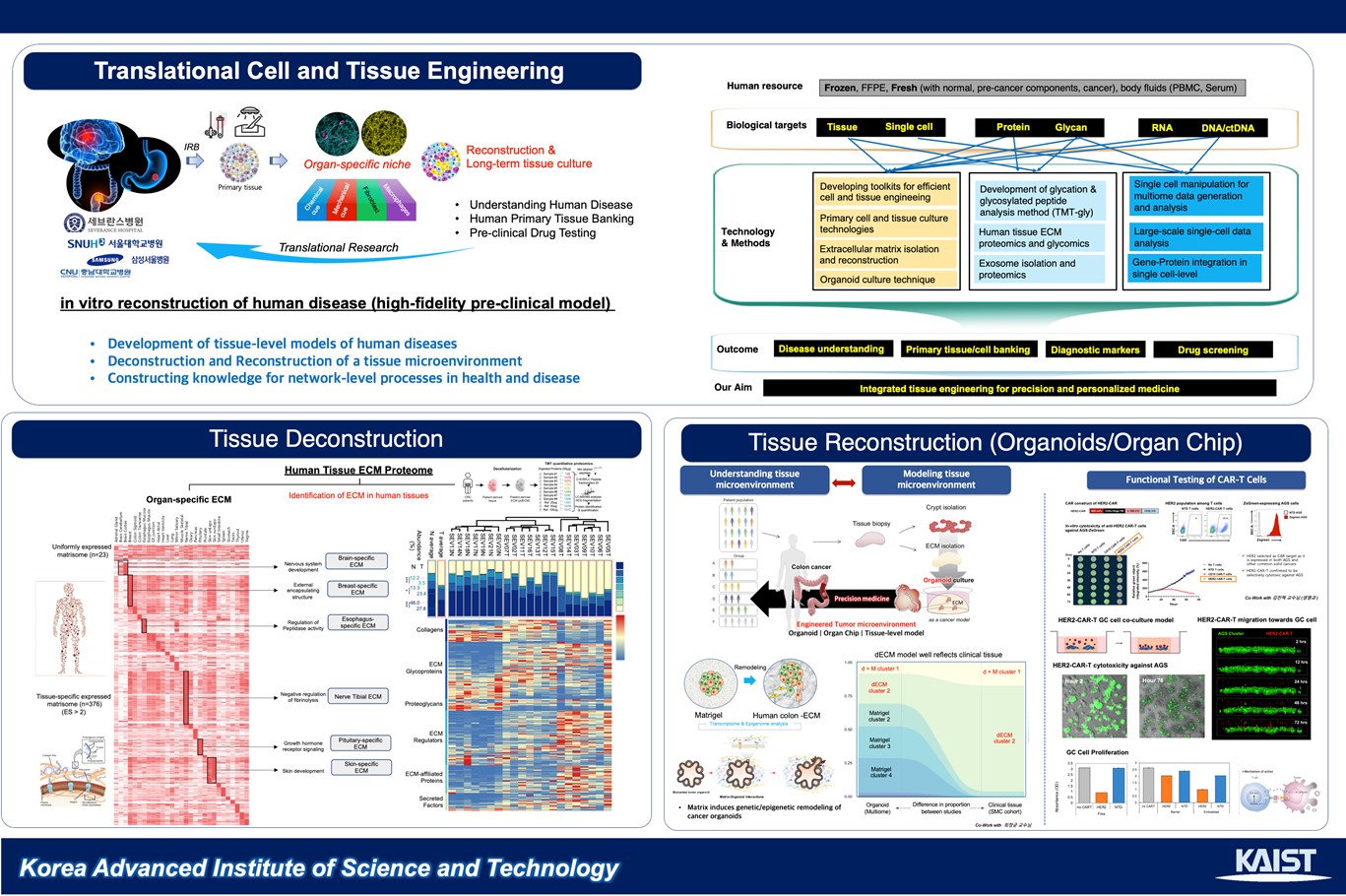
3) Extracellular Matrix Engineering (Matrisome)
Extracellular matrix (ECM) of the microenvironment is a 3D architecture that consists of proteins, minerals, and other macromolecules that act as a structural support and chemical reservoir to neighboring cells. Simply, it is all of the non-cellular components in tissue. Commonly, fibrous architecture is formed by collagens, fibrinogens, hydroxyapatite, and other materials, but each organ and tissue have different ratios and compositions of such ECM components. Hence, it is important to consider the quantity of each ECM protein in designing a more accurate in vitro tumor microenvironment. Decellularization is a method that removes cellular components from tissues while leaving intact the tissue-specific biochemical profile of ECM. Current in vitro hydrogel culture models, such as collagen type-1 and matrigel, can only partially mimic physiological properties of native tissue microenvironment. To better reconstitute tissue-specific microenvironments, decellularization techniques supply the tissue-specific ECM molecules and growth factors that affect various cell-matrix interactions and pathologies in in vitro culture systems using cancer cell lines and tumor organoid models. Further, the analysis of decellularized ECM (dECM) through mass spectrometry (proteomics) and RNA-sequencing of organoids cultured in dECM versus other hydrogel materials help us determine the role of the ECM in the cellular context.
4) Mechanotransduction
Among many extracellular cues, mechanical stimuli are especially important in the regulation of cell behavior in cell adhesion, proliferation, and cell cycle regulation. Mechanotransduction, or the translation of mechanical signals to biological response, is hypothesized to have a role in the progression of cancer, as it is often observed that tissues that surround the tumor area are stiffer than normal tissues. The stiffened extracellular matrix conveys biophysical cues that are transmitted to the nucleus via cytoskeletal proteins. Mechanosensors and mechanotransducers such as Yes-associated protein, are examined in detail. At BiMiL, we highlight the importance of the mechanical characteristics of the tumor microenvironment as well as the biochemical factors in constructing a tumor model. We continue to aim to uncover the role of mechanotransduction in tumor tissues and the clinical impact of the balance between mechanical and chemical signals in cancer progression.
Key Achievements
- 1. P. Kim, M. Abkarian, H. A. Stone*, “Hierarchical folding of elastic membranes under biaxial compressive stress”, Nat. Mater. 10, 952–957 (2011)
- 2. H. N. Kim*, K.-J. Jang, J.-Y. Shin, D. Kang, S. M. Kim, I. Koh, Y. Hong, S. Jang, M. S. Kim, B.-S. Kim, H. E. Jeong, N. L. Jeon, P. Kim*, and K.-Y. Suh, "Artificial Slanted Nanocilia Array as a Mechanotransducer for Controlling Cell Polarity", ACS Nano, 11, 730, (2017)
- 3. J. Cha, H. Kim, N. Hwang, P. Kim*, "Mild Reduction of the Cancer Cell Surface as an Anti-invasion Treatment", ACS Applied Materials & Interfaces, 10, 35676 (2018)
- 4. J. Cha, P. Kim*, "Time series assessment of the effects of hypoxic stress on glioma tumorsphere development within engineered microscale niches", Biomaterials, 194, 171 (2019)
- 5. M. Jang, G. Choi, Y. Y. Choi, J. E. Lee, D. H. Han, H. Lee, J.-H. Cheong, P. Kim*, "Extracellular vesicles (EVs)-bioadhesives nanoaggregates for microRNA-based cancer diagnosis", 11, 79, NPG Asia Materials (2019)