


Broad research interests of magnetic resonance imaging (MRI) laboratory at Korea Advanced Institute of Science and Technology (KAIST) lie in (i) developing new MRI techniques of image acquisition, processing, and analysis, (ii) unveiling the inter-regional brain communication mechanism, (iii) applying machine learning algorithms to MRI databases and MRI artifact suppression, and (iv) developing intraoperative MRI devices, These technical developments can make big differences in understanding baseline and functional brain physiology, and also diagnosis / prognosis / treatment of our body.
Research Area
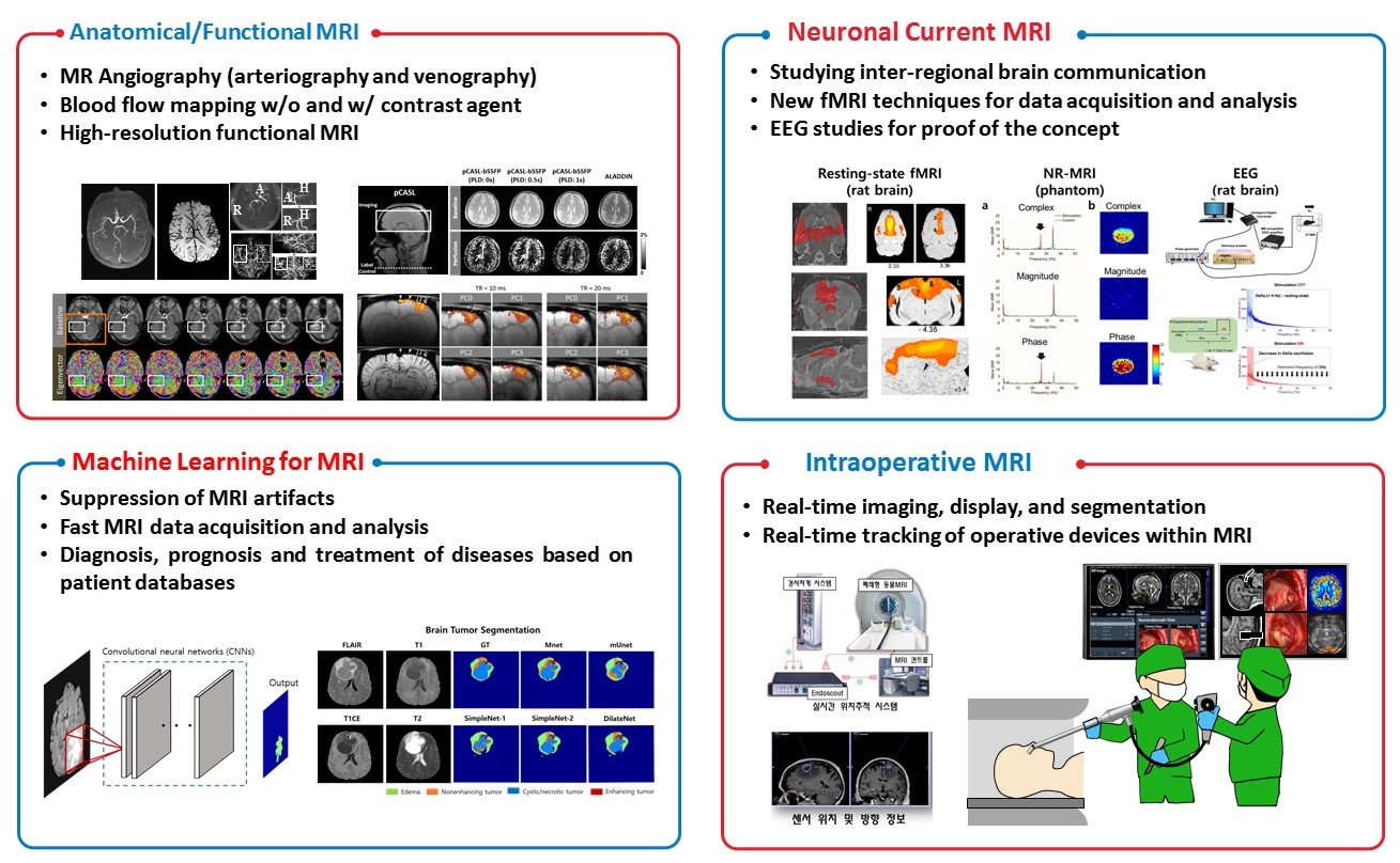
1) Anatomical/Physiological/Functional MRI
Noninvasive visualization of blood vessels, blood flow, and blood oxygenation level dependence is an important step toward understanding sources of functional brain signals and brain diseases. We are developing methods to image both arteries and veins within a single MRI acquisition without compromising their image qualities, map blood flow in brain, retina, and kidney noninvasively, and map brain function with high spatial resolutions. These techniques have been used to understand signal sources of high-resolution functional MRI and also vascular origin of brain diseases such as brain tumors and stroke
2) Neuronal Current MRI
Our brain requires coherent information flow for inter-regional communication. However, it is still not clear how a neuronal group selects its communication target. We are trying to develop a new fMRI technique that enables us to detect neuronal currents directly rather than the conventional fMRI relying on secondary hemodynamic responses. We try to use the new fMRI technique as well as systems biological approaches for better understanding of the inter-regional brain communication mechanism and use EEG to prove the concept.
3) Machine Learning for Biomedical Imaging
Machine learning has been of interest to the many research areas and one of the hottest applications is biomedical imaging. We are trying to develop and apply machine learning algorithms for MRI artifacts suppression, denoising, fast MRI data acquisition and analysis, and diagnosis/prognosis/treatment monitoring of various diseases
4) Intraoperative MRI
Intraoperative MRI is application of MR imaging during or in-between medical treatments. We are developing system for real-time imaging, display, and segmentation as well as real-time tracking of operative devices within MRI
5) Biomedical Applications
We have tried to integrate all these imaging techniques for multi-parametric assessment of various brain diseases. We believe combination of the various aspects of the brain would provide much more accurate information than conventional approaches. Also we expect this approach can stimulate interdisciplinary collaborative studies involving engineers, neuroscientists, and clinicians.
-
1. Jun-Hee Kim#, Jae-Geun Im#, and Sung-Hong Park, Measurement of CSF Pulsation from EPI-based Human fMRI, NeuroImage, 2022;257:119293
-
2. Ji Hoon Ahn#, Hyunsoo Cho#, Jun-Hee Kim#, Shin Heun Kim, Je-Seok Ham, Sang Heon Suh, Seon Pyo Hong, Joo-Hye Song, Young-Kwon Hong, Yong Jeong, Sung-Hong Park, and Gou Young Koh, Meningeal lymphatic vessels at the skull base drain cerebrospinal fluid, Nature, 2019, 572(7767):62-66
-
3. Won-Joon Do, Seung-Hong Choi, and Sung-Hong Park. Simultaneous Variable-slab Dual-echo TOF MR Angiography and Susceptibility-Weighted Imaging, IEEE Transactions on Medical Imaging, 2018;37(7):1632-1640
-
4. Ki Hwan Kim, Seung Hong Choi, and Sung-Hong Park. Improving Arterial Spin Labeling by using Deep Learning, Radiology, 2018;287(2):658-666
-
5. Ki Hwan Kim, Won-Joon Do, and Sung-Hong Park. Improving Resolution of MR Images with an Adversarial Network Incorporating Images with Different Contrast, Medical Physics, 2018;45(7):3120-3131
- 6. Ji Hoon Ahn#, Hyunsoo Cho#, Jun-Hee Kim#, Shin Heun Kim, Je-Seok Ham, Sang Heon Suh, Seon Pyo Hong, Joo-Hye Song, Young-Kwon Hong, Yong Jeong, Sung-Hong Park*, and Gou Young Koh*, Meningeal lymphatic vessels at the skull base drain cerebrospinal fluid, Nature, 2019, 572(7767):62-66,
- 7. Ki Hwan Kim, Seung Hong Choi, and Sung-Hong Park. Improving Arterial Spin Labeling by using Deep Learning, Radiology, 2018;287(2):658-666
- 8. Won-Joon Do, Seung Hong Choi, and Sung-Hong Park. Simultaneous variable-slab dual-echo TOF MR angiography and susceptibility-weighted imaging, IEEE Transactions on medical imaging, 2018;37(7):1632-1640.
- 9. Paul Kyu Han, Seung Hong Choi, and Sung-Hong Park. Investigation of Control Scans in Pseudo-Continuous Arterial Spin Labeling (pCASL): Strategies for Improving Sensitivity and Reliability of pCASL, Magnetic Resonance in Medicine, 2017;78(3):917-929
- 10. Sung-Hong Park, Tae Kim, Ping Wang, and Seong-Gi Kim. Sensitivity and specificity of high-resolution balanced steady state free precession fMRI at high field of 9.4T. NeuroImage 2011, Sep;58(1):168-176.