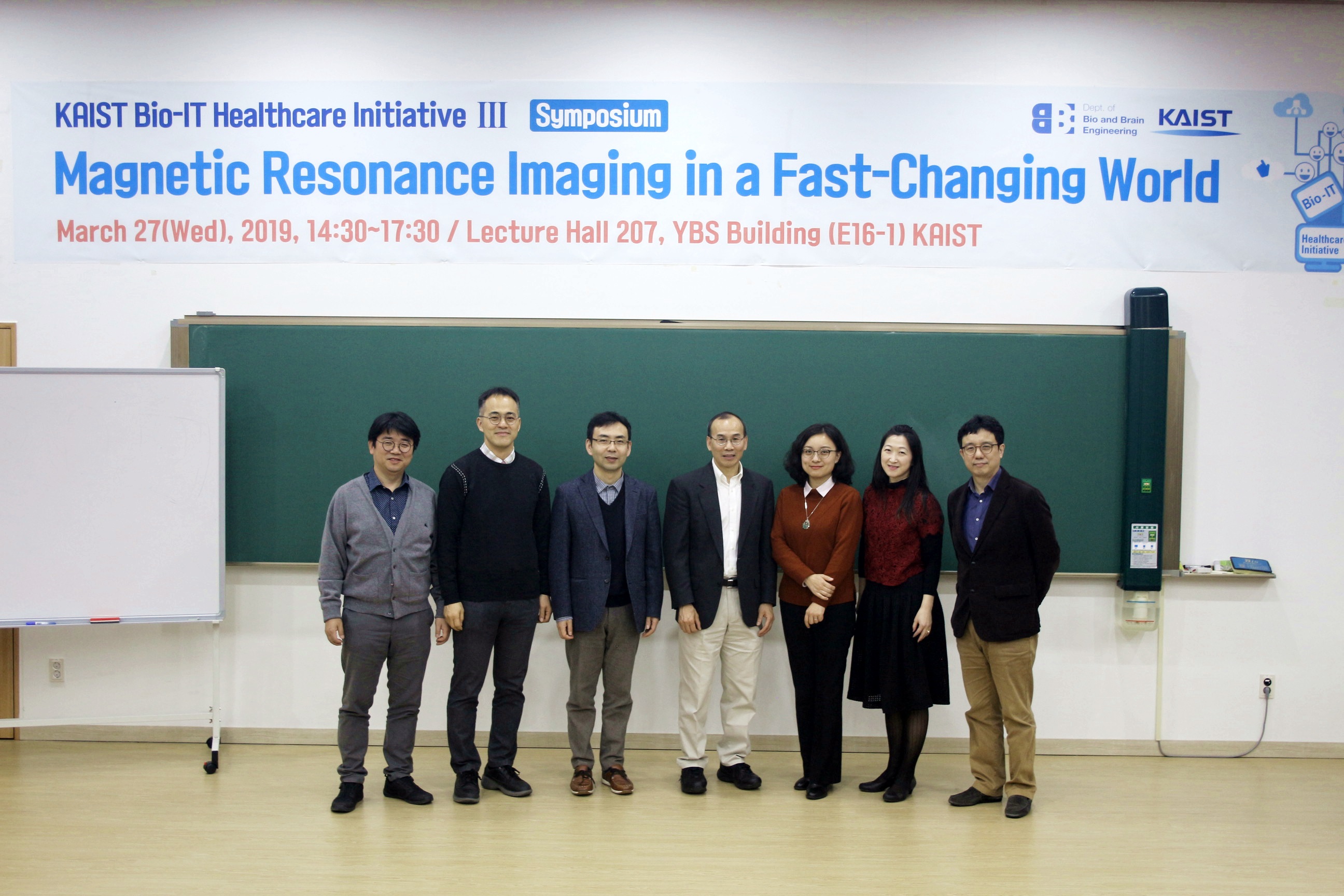
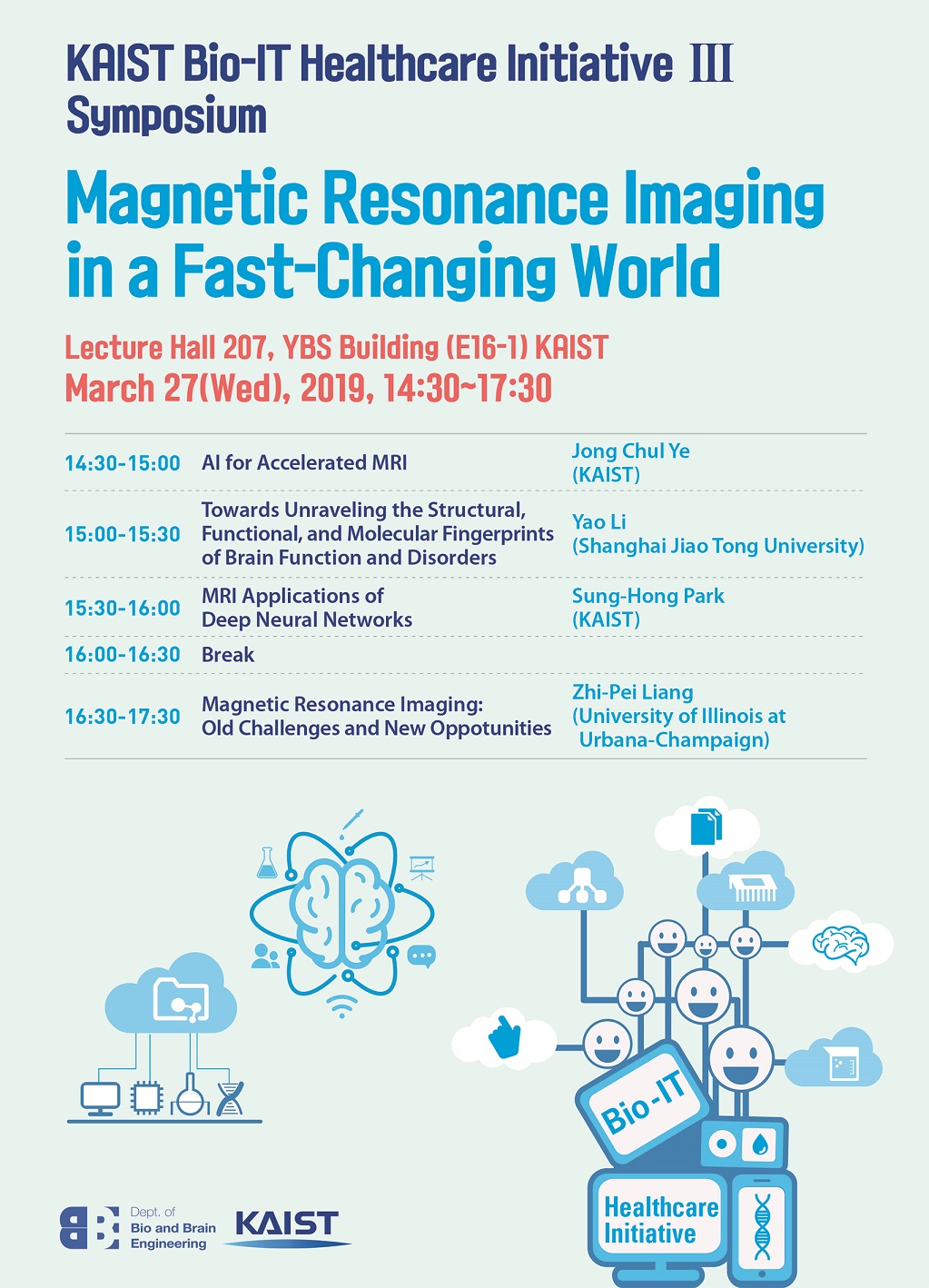
3/27일(수) 14:30~17:30까지 KAIST Bio-IT Healthcare Initiative III -Globalization 사업의 일환으로 심포지움 “Magnetic Resonance Imaging in a Fast-Changing World” 가 개최되었습니다.
본 심포지움에서는 해외석학들과 함께 MRI 연구분야가 4차산업혁명 시대에 어떻게 변화되고 있는지 최신 기술들과 그를 활용한 연구들을 통해 짚어보고 앞으로 나아갈 방향에 대한 활발한 논의가 이루어졌습니다.
Invited Spearker1: Professor Zhi-Pei Liang ( Franklin W. Woeltge Professor, Department of Electrical and Computer Engineering
Co-chair, Integrative Imaging Theme, Beckman Institute, University of Illinois at Urbana-Champaign)
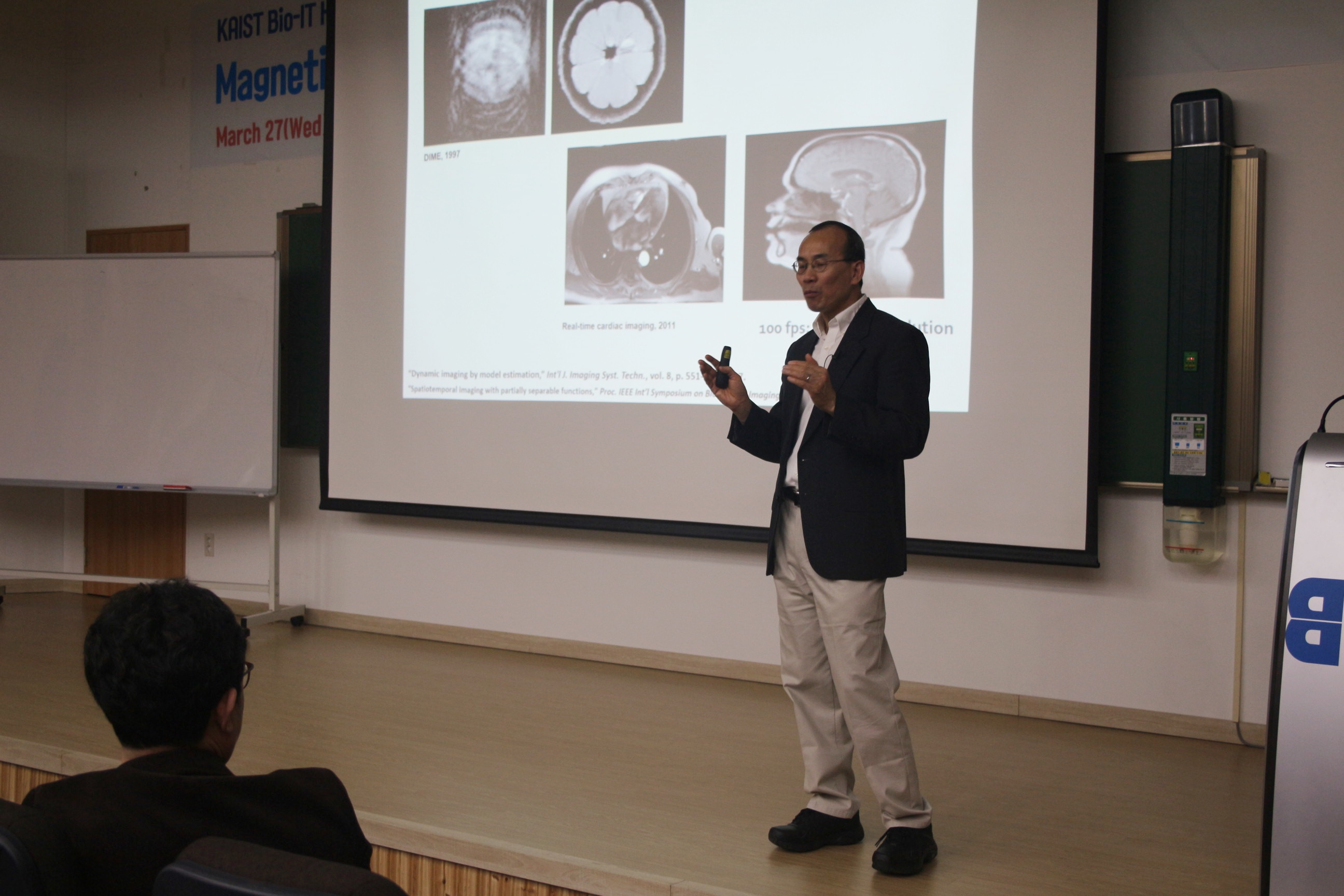
* Title: Magnetic Resonance Imaging: Old Challenges and New Opportunities
* Abstract: Nuclear magnetic resonance was first observed in bulk matter by Edward Purcell's group at Harvard and Felix Bloch's group at Stanford in 1946. Magnetic resonance imaging (MRI) was first proposed by Paul Lauterbur in 1972. Since then, MRI has revolutionized medicine and biology. With the ever increasing use of MRI in biomedical research and clinical applications, MRI is entering a new era of technology development and practical applications. This talk will give an overview of MRI, discussing some of the long-standing technical challenges and new opportunities in the context of the new advances in computational imaging.
Invited Speaker2: Professor Yao Li ( Institute for Medical Technoloty, School of Biomedical Engineering, Shanghai Jiao Tong University)
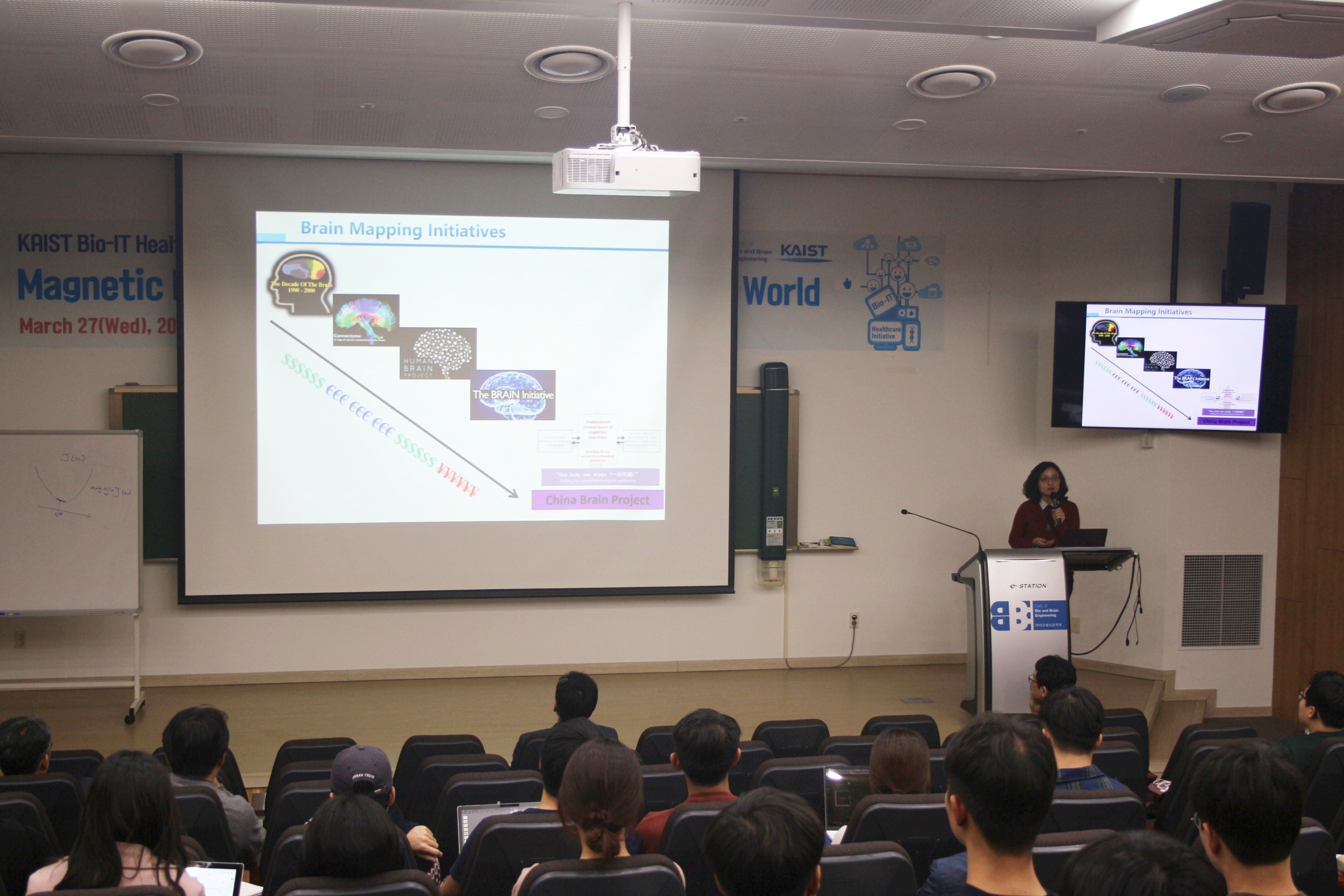
*Title: Towards Unraveling the Structural, Functional, and Molecular Fingerprints of Brain Function and Disorders
*Abstract: Brain mapping is one of the most exciting frontiers of contemporary science. It provides this generation of scientists the opportunity to make major advances on a historic question: how the brain works and what goes wrong when it is injured or diseased. Magnetic resonance (MR)-based neuroimaging techniques have been widely used for brain research because of their noninvasiveness and multimodal capabilities. Exploiting the rich information contents of MR signals, we can obtain structural, functional and metabolic information of the brain. MR spectroscopic imaging (MRSI) is a beautiful integration of MR spectroscopy and MR imaging, which can provide metabolic status of tissues in vivo without exogenous molecular labels. But its clinical applications have been limited due to long scan time and poor spatial resolution. In this talk, I’ll show a new capability for rapid high-resolution metabolic imaging by using a recently developed subspace-based imaging technique called SPICE (SPectroscopic Imaging by exploiting spatiospectral CorrElation). In a 5-min scan, we can acquire metabolic maps from the whole brain at a nominal spatial resolution of 2.0 x 3 x 3 mm3. We have successfully performed SPICE scans on stroke patients, traumatic brain injury (TBI) patients and brain tumor patients. Our experimental study yielded very encouraging results and showed that ultrahigh-resolution MRSI can capture neurometabolic alterations induced by stroke, TBI and brain tumors effectively.